Area on a Bone Where Two or More Bones Articulate (Move in Relation to Each Other)
Introduction [edit | edit source]
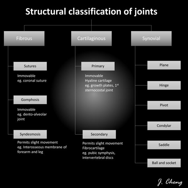
Joints, also known as articulations, are a form of connection between bones. They provide stability to the skeletal system as well as allowing for specialized movement.
Joints can be classified:
- Histologically, on the dominant type of connective tissue. ie fibrous, cartilaginous, and synovial.
- Functionally, based on the amount of movement permitted. ie synarthrosis (immovable), amphiarthrosis (slightly moveable), and diarthrosis (freely moveable)[1].
Generally speaking, the greater the range of movement, the higher the risk of injury because the strength of the joint is reduced
The two classification schemes correlate:
- Synarthroses are fibrous joints
- Amphiarthroses are cartilaginous joints
- Diarthroses are synovial joints
The 5 minute video outlines the basics.
Fibrous Joints [edit | edit source]
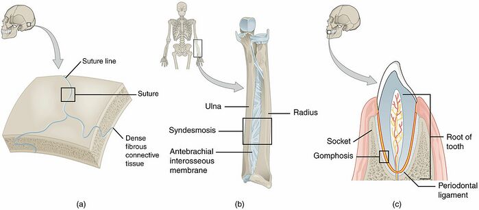
In fibrous joints (synarthrodial joint) the bones are joined by fibrous tissue, namely dense fibrous connective tissue, and no joint cavity is present. The amount of movement allowed depends on the length of the connective tissue fibers uniting the bones. Although a few are slightly movable, most fibrous joints are immovable.
The three types of fibrous joints are sutures, syndesmoses, and gomphoses.
- Sutures are immobile joints in the cranium. The plate-like bones of the skull are slightly mobile at birth because of the connective tissue between them, termed fontanelles. This initial flexibility allows the infant's head to get through the birth canal at delivery and permits the enlargement of the brain after birth. As the skull enlarges, the fontanelles reduce to a narrow layer of fibrous connective tissue that suture the bony plates together. Eventually, cranial sutures ossify- the two adjacent plates fuse to form one bone (termed synostosis).
- Gomphoses are the immobile joints between the teeth and their sockets in the mandible and maxillae. The periodontal ligament is the fibrous tissue that connects the tooth to the socket.
- Syndesmoses are slightly movable joints (amphiarthroses). In syndesmosis joints, the two bones are held together by an interosseous membrane. Eg Middle Tibiofibular Joint, a fibrous joint formed by the interosseus membrane connecting the shafts of the tibia and the fibula[1].
Cartilaginous Joints [edit | edit source]
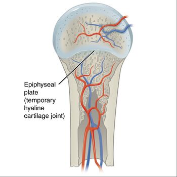
Cartilaginous joints are a type of joint where the bones are entirely joined by cartilage, either hyaline cartilage or fibrocartilage. These joints generally allow more movement than fibrous joints but less movement than synovial joints.
- Primary cartilaginous joints: These cartilaginous joints are composed entirely of hyaline cartilage and are known as synchondroses. Most exist between ossification centres of developing bones and are absent in the mature skeleton, but a few persist in adults. eg First Sternocostal Joint, between first rib and manubrium (all other sternocostal joints are plane synovial joints); Growth plates. Image 3: synchondroses eg. growth plate
- The secondary cartilaginous joint, also known as symphysis, may involve either hyaline or fibrocartilage. These joints are slightly mobile (amphiarthroses). eg The pubic symphysis: Intervertebral discs[2].
Synovial Joints [edit | edit source]
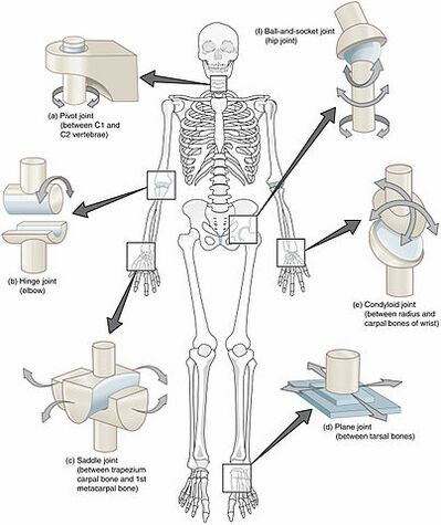
The primary purpose of the synovial joint is to prevent friction between the articulating bones of the joint cavity. While all synovial joints are diarthroses, the extent of movement varies among different subtypes and is often limited by the ligaments that connect the bones. Nearly all joints of the limbs and most joints of the body fall into this class.
A key structural characteristic for a synovial joint that is not seen at fibrous or cartilaginous joints is the presence of a joint cavity. The joint cavity contains synovial fluid, secreted by the synovial membrane (synovium), which lines the articular capsule. This fluid-filled space is the site at which the articulating surfaces of the bones contact each other. Hyaline cartilage forms the articular cartilage, covering the entire articulating surface of each bone. The articular cartilage and the synovial membrane are continuous. A few synovial joints of the body have a fibrocartilage structure located between the articulating bones. This is called an articular disc, which is generally small and oval-shaped, or a meniscus, which is larger and C-shaped.[1]
See Synovial Joint Link for full explaination.
Physiotherapy [edit | edit source]
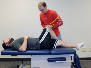
Physiotherapists are qualified health care professionals who are experienced at assessing joints of the human body. See links to below conditions for some examples.
- Arthritis – inflammation that causes stiffness and pain in the joints eg rheumatoid arthritis or gout, or degeneration (osteoarthritis)
- Bursitis – inflammation of the bursae (fluid-filled sacs that cushion and pad bones)
- Tendonitis – inflammation, irritation and swelling of a tendon that is attached to the joint.
- Injury – including strain or sprain of a ligament or nearby tendon or muscle, or bone fracture.[3]
References [edit | edit source]
- ↑ 1.0 1.1 1.2 Juneja P, Hubbard JB. Anatomy, Joints.Available: https://www.ncbi.nlm.nih.gov/books/NBK507893/ (accessed 21.6.2021)
- ↑ Radiopedia Cartilaginous Joints Available: https://radiopaedia.org/articles/cartilaginous-joints(accessed 21.6.2021)
- ↑ Better Health Joints Available: https://www.betterhealth.vic.gov.au/health/conditionsandtreatments/joints#joint-conditions (accessed 21.6.2021)
Area on a Bone Where Two or More Bones Articulate (Move in Relation to Each Other)
Source: https://www.physio-pedia.com/Joint_Classification
0 Response to "Area on a Bone Where Two or More Bones Articulate (Move in Relation to Each Other)"
Post a Comment